Please use this identifier to cite or link to this item:
https://ahro.austin.org.au/austinjspui/handle/1/9474| Title: | Occipital epilepsies: identification of specific and newly recognized syndromes. | Austin Authors: | Taylor, Isabella;Scheffer, Ingrid E ;Berkovic, Samuel F | Affiliation: | Epilepsy Research Institute, University of Melbourne, Austin and Repatriation Medical Centre, Heidelberg West, Victoria, Australia | Issue Date: | 1-Apr-2003 | Publication information: | Brain : A Journal of Neurology; 126(Pt 4): 753-69 | Abstract: | Occipital epilepsies often elude diagnosis as they frequently masquerade as other seizure syndromes. Visual hallucinations are the key clinical symptoms indicating an occipital focus, but may be difficult to elicit on history, especially from children, and are not always present. When visual symptoms are not prominent, the seizure semiology and scalp EEG may lead the clinician away from considering an occipital focus, as they often reflect seizure propagation rather than seizure origin. Clinical and neuroimaging advances have led to the recognition of many new occipital epilepsy syndromes, which generally present in childhood or adolescence. Major groups include malformations of cortical development [focal cortical dysplasia, periventricular heterotopia (PVH), subcortical band heterotopia (SBH), polymicrogyria], vascular (including epilepsy with bilateral occipital calcifications often associated with coeliac disease), metabolic and the emerging idiopathic occipital epilepsies. The idiopathic occipital epilepsies now comprise three identifiable electroclinical syndromes of childhood and adolescence, the biological inter-relationships and overlap with idiopathic generalized epilepsies of which are discussed here. We emphasize the clues to recognition of specific occipital epilepsies, some of which now have specific treatments. Where medical therapy is ineffective, occipital corticectomy should be considered. Emerging evidence suggests that some syndromes have a good surgical outcome, and the consequences to visual function may be less severe than anticipated. | Gov't Doc #: | 12615636 | URI: | https://ahro.austin.org.au/austinjspui/handle/1/9474 | Journal: | Brain | URL: | https://pubmed.ncbi.nlm.nih.gov/12615636 | Type: | Journal Article | Subjects: | Cerebral Cortex.abnormalities Diagnosis, Differential Electroencephalography Epilepsy.diagnosis.etiology Hallucinations.etiology Humans Magnetic Resonance Imaging.methods Migraine Disorders.diagnosis Occipital Lobe Syndrome Tomography, X-Ray Computed.methods |
| Appears in Collections: | Journal articles |
Files in This Item:
| File | Description | Size | Format | |
|---|---|---|---|---|
| 12615636.pdf | 367.48 kB | Adobe PDF | 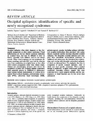 View/Open |
Page view(s)
46
checked on Dec 20, 2024
Download(s)
136
checked on Dec 20, 2024

Google ScholarTM
Check
Items in AHRO are protected by copyright, with all rights reserved, unless otherwise indicated.