Please use this identifier to cite or link to this item:
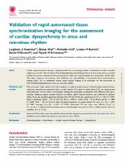
https://ahro.austin.org.au/austinjspui/handle/1/11188| Title: | Validation of rapid automated tissue synchronization imaging for the assessment of cardiac dyssynchrony in sinus and non-sinus rhythm. | Austin Authors: | Kearney, Leighton G ;Wai, Bryan;Ord, Michelle;Burrell, Louise M ;O'Donnell, David ;Srivastava, Piyush M | Affiliation: | The University of Melbourne, Austin Health and Northern Health, Melbourne, Australia | Issue Date: | 1-Feb-2011 | Publication information: | Europace : European Pacing, Arrhythmias, and Cardiac Electrophysiology : Journal of the Working Groups On Cardiac Pacing, Arrhythmias, and Cardiac Cellular Electrophysiology of the European Society of Cardiology; 13(2): 270-6 | Abstract: | Cardiac resynchronization therapy is showing benefits for an increasing number of indications but fails to predict response in up to 20-30% of subjects. Echocardiographically assessed dyssynchrony has been proposed as a potential stratifier but current methods are time-consuming and suffer poor reproducibility, thus limiting their clinical utility. This study compared the accuracy, time efficiency, and reproducibility of automated tissue synchronization imaging (Auto TSI) vs. established manual tissue velocity imaging (TVI) techniques for the assessment of intra-ventricular dyssynchrony in sinus and non-sinus rhythm.Fifty consecutive stable systolic heart failure patients on optimal guideline-based medical therapy underwent intra-ventricular dyssynchrony assessment [time to peak velocity (Ts), septal to lateral delay (SLD), and dyssynchrony index (DI)] with TVI and Auto TSI techniques, enabling the assessment of agreement, time efficiency, and reproducibility. Statistical analyses included Pearson's correlation, Bland-Altman's statistics, and coefficient of reproducibility. There was excellent agreement between Auto TSI and TVI for the measurement of Ts [r=0.92, P<0.001, limits of agreement (LOA): -27.3 to 56.5 ms], SLD (r=0.94, P<0.001, LOA: -41 to 49 ms), and DI (r=0.89, P<0.001, LOA: -12.2 to 12.6 ms) which persisted irrespective of cardiac rhythm [Ts: sinus (n=32) r=0.93, P<0.001; non-sinus (n=18) r=0.91, P<0.001]. Automated TSI was more time efficient (3±1 vs. 14±2 min, P<0.001) and demonstrated superior reproducibility: intra-observer (5.5 vs. 9.6%) and inter-observer variability (9.5 vs. 13.4%).Automated TSI enables rapid, reproducible intra-ventricular dyssynchrony assessment and overcomes some of the limitations of conventional techniques in sinus and non-sinus rhythm. | Gov't Doc #: | 21252196 | URI: | https://ahro.austin.org.au/austinjspui/handle/1/11188 | DOI: | 10.1093/europace/euq442 | Journal: | Europace : European pacing, arrhythmias, and cardiac electrophysiology : journal of the working groups on cardiac pacing, arrhythmias, and cardiac cellular electrophysiology of the European Society of Cardiology | URL: | https://pubmed.ncbi.nlm.nih.gov/21252196 | Type: | Journal Article | Subjects: | Cardiac Resynchronization Therapy Diagnostic Imaging.methods Echocardiography, Doppler, Color.methods Heart Failure.physiopathology.therapy Humans Observer Variation Reproducibility of Results Sick Sinus Syndrome.physiopathology Treatment Outcome |
| Appears in Collections: | Journal articles |
Files in This Item:
| File | Description | Size | Format | |
|---|---|---|---|---|
| 21252196.pdf | 251.58 kB | Adobe PDF |  View/Open |
Page view(s)
58
checked on Apr 17, 2025
Download(s)
112
checked on Apr 17, 2025
Google ScholarTM
Check
Items in AHRO are protected by copyright, with all rights reserved, unless otherwise indicated.
